Location:Home >> Detail
Med One. 2016; 1(1): 1; https://doi.org/10.20900/mo.20160001
 ,
Feipeng Wang2,
Lei Wang3,
Liangxiang Chen4,
Yuhong Shi5
,
Feipeng Wang2,
Lei Wang3,
Liangxiang Chen4,
Yuhong Shi5
1 Gynaecology Department, The Tumor Hospital of Shaanxi Province, 710061, China
2 Gynaecology Department, Xianyang Central Hospital, Shaanxi Province, 712000, China
3 Gynaecology Department, First People's Hospital of Xianyang, Shaanxi Province, 712000, China
4 Gynaecology Department, Hunan Province Maternal and Child Health Care Hospital, China
5 Department of Surgery, University of Maryland School of Medicine, Baltimore, MD 21221, USA
*Correspondence: Xuemei Yang.
Background: Interleukin-37 (IL-37) is member 7 of the IL-1 family and inhibits pro-inflammatory factor activities. Cervical carcinoma is a malignant tumor associated with inflammation. The roles of IL-37 in the occurrence and progression of cervical cancer have not been elucidated. This study investigated the roles of IL-37 in the proliferation, survival, invasion, and migration of cervical cancer cells.
Methods: Cell viability in IL-37-treated HeLa cells was measured using MTT assay. Cell cycle and apoptosis were analyzed with flow cytometry. The invasion, and migration, of cells was tested using the Transwell approach. STAT3, MMP-2, and MMP-9 protein expression was measured with Western blot.
Results: In HeLa cells, L-37 treatment significantly reduced viability, increased the percentage of G0/G1 cells, and induced cell apoptosis in a dose- and time-dependent manner (p < 0.05). For cervical carcinoma cells, IL-37 treatment significantly inhibited their invasion and migration, and lowered STAT3, MMP-2, and MMP-9 protein expression (p < 0.05).
Conclusion: IL-37 functions as a tumor suppressor in cervical cancer cells by inhibiting cell proliferation, cell cycling, and invasion/migration possibly through hindering the STAT3-MMP-2/MMP-9 signaling.
Cervical cancer is the most common female reproductive system malignancy. Its incidence and mortality are rapidly increasing worldwide [1]. Cervical carcinoma is an infectious disease of the cervix. Human papillomavirus (HPV) infection is thought to be the primary cause cervical carcinoma. Herpes virus II, Neisseria gonorrhoeae, Chlamydia trachomatis, and bacterial vaginosis have also been associated with cervical inflammation [2]. Inflammation plays a major role in cervical cancer occurrence and progression [3]. Studies of the molecular mechanisms of cervical cancer occurrence and progression have revealed that cervical tumor cells secrete large amounts of inflammatory factors, which could affect cellular signaling pathways, subsequently inhibit immune recognition of tumor cells, and affect tumor cell proliferation, invasion, and metastasis [4]. The roles of various inflammatory factors in the malignant phenotype of cervical cancer are not yet fully understood.
Interleukin-1 (IL-1) is an 11-member family of cytokines that regulate inflammation [5]. IL-37 is member 7 of the IL-1 family and was first discovered in 2000 by sequence alignment. It is was located on chromosome 2 with 5 different splicing subtypes [6]. Studies have found that IL-37 plays an important role in multiple inflammation-associated diseases [7, 8]. Only a few studies have reported on the biological roles of IL-37 in cancer. IL-37 was found to mediate renal cell carcinoma [9] and hepatocellular carcinoma anti-tumor activities [10]. One study reports, IL-37 as suppressing cell proliferation and cervical cancer cell invasion by downregulation of STAT3 phosphorylation [11]. This study explores the roles of IL-37 in cell proliferation, survival, and metastasis, as well as the associated signaling, in cervical cancer cells.
Recombinant human IL-37 (RhIL-37) was obtained from ProSpec Company. Transwell chambers were obtained from Milipore. Rabbit anti-STAT3 monoclonal antibodies, rabbit anti-phosphorylated STAT3 antibodies, and rabbit anti-MMP-2 and MMP-9 antibodies were purchased from Abcam. Mouse, anti-human β-actin, HRP-conjugated goat anti-mouse IgG, and HRP-conjugated goat anti-rabbit IgG antibodies were purchased from Dingguo Company.
2.2 Cell cultureThe human cervical cancer HeLa cell line was provided by the Chinese Cell Culture Center (Wuhan, China). The cells were cultured in either a RPMI 1640 medium, or a DMEM medium (Gibco, US) containing 2 mmol/L l-glutamine, 100 U penicillin, 100 μg/ml streptomycin, and 2 %, or 10 %, fetal bovine serum (Gibco,US) at 37°C, 5 % CO2.
2.3 MTT assayCells were cultured with IL-37 at final concentrations of 0 ng/mL, 50 ng/mL, 100 ng/mL, 200 ng/mL, and 400 ng/mL for 24 hrs, 48 hrs, and 72 hrs. Twenty μL of an MTT solution (5 mg/mL) was added to each well and the cells were incubated for 4 hrs. Next, 150μL of DMSO was added to each well and the cells were incubated for 10 min under gentle agitation. Absorbance values were then measured at 570 nm.
2.4 Flow cytometryHeLa cells at the log phase were trypsinized and seeded at 2 × 10⁶ cell/dish into 10 cm dishes. After incubation in 2 % fetal bovine serum for 24 hrs, the culture medium was changed to DMEM which contained 10 % fetal bovine serum. Different concentrations of IL-37 (final concentration: 0 ng/mL, 50 ng/mL, 100 ng/mL, and 200 ng/mL) were added to cells and continuously cultured for different periods of time. Cells were then collected and centrifuged at 1200r/min for 5 min to remove supernatants. After washing with 1 × PBS, cells were fixed in absolute ethanol overnight at 4°C. Propidium iodide (PI) dye was added, and cells were incubated at 4°C for 30 min followed by flow cytometry assay.
2.5 Cell invasion and migration assayTranswell chambers were coated with matrigel overnight at 4°C and the basal membrane was hydrated with serum-free culture medium for 1h at 37°C to perform an invasion assay. Treated HeLa cells were seeded into the upper chamber of the Transwell chamber. RPMI 1640 medium was added into the lower chamber. Twenty-four hours later, the membranes were stained using Giemsa dye and cells were numbered by counting using an inverted microscope. The artificial basal membrane was excluded from the invasion assay in order to perform a migration essay.
2.6 Western blotHeLa cells were cultured in 10 cm dished with DMEM medium containing 10 % fetal bovine serum. Cells at the log-phase were treated with 200 ng/ml of IL-37 for 48 hrs. Cells were then homogenized and total protein was loaded on an 8 % SDS-PAGE gel (40 μg per lane). After transferring proteins to the PVDF membrane, the membranes were blocked with 1 × TBST buffer (1 × TBS buffer with 0.1 % Tween-20) containing 5 % non-fat milk for 1 hr at room temperature (RT), followed by incubation with a primary antibody (1:2000 dilution for phospho-STAT3, total MMP-2, MMP-9, and β-actin) overnight at 4°C and further incubation for 30 min at RT. After washing with 1 × TBST, membranes were incubated with an HRP-conjugated secondary antibody (1:2000 dilution) for 1 hr at RT. After washing with 1 × TBST for 3 times, immune reaction was visualized by incubation with chromogenic substrates and exposure to X-ray films. Band optical density was scanned and analyzed using a Quantity One software package.
2.7 Data analysisData were analyzed using SPSS17.0 statistical software and presented as mean±standard deviation (SD). Comparison between the two groups was performed using a student’s t-test. Statistical significance was defined as p < 0.05.
HeLa cells were cultured with 10 % FCS and treated with different concentrations of IL-37 (0 ng/mL, 50 ng/mL, 100 ng/mL, 200 ng/mL, and 400 ng/mL) for 24 hrs, 48 hrs, and 72 hrs. An MTT assay showed that IL-37 (50-400 ng/mL) significantly reduced cell viability at all time points (p < 0.05) in a dose- and time-dependent manner (Table 1).
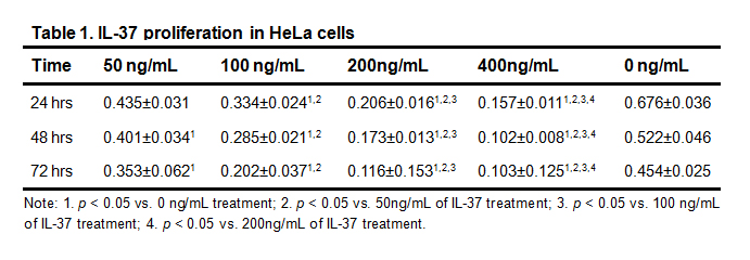 Table 1. IL-37 proliferation in HeLa cells
Note: 1. p < 0.05 vs. 0 ng/mL treatment; 2. p < 0.05 vs. 50ng/mL of IL-37 treatment; 3. p < 0.05 vs. 100 ng/mL of IL-37 treatment; 4. p < 0.05 vs. 200ng/mL of IL-37 treatment.
Flow cytometry was used to detect apoptosis and analyze the cell cycle of IL-37-treated HeLa cells. Treatment with 50 ng/mL, 100 ng/mL, 200 ng/mL, and 400 ng/mL of IL-37 for 48 hrs significantly increased HeLa cells numbers in the G0/G1 phase, but decreased HeLa cell numbers in the S phase, as compared to cells treated with 0 ng/mL of IL-37 (p < 0.05) in a concentration-dependent way. IL-37 significantly increased the percentage of apoptotic HeLa cells (p < 0.05) in a concentration-dependent manner (Table 2).
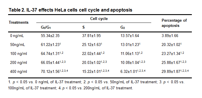 Table 2. IL-37 effects HeLa cells cell cycle and apoptosis
1. p < 0.05 vs. 0 ng/mL of IL-37 treatment; 2. p < 0.05 vs. 50ng/mL of IL-37 treatment; 3. p < 0.05 vs. 100ng/mL of IL-37 treatment; 4. p < 0.05 vs. 200ng/mL of IL-37 treatment.
A Transwell assay was used to analyze the invasion and migration ability of IL-37-treated HeLa cells. Results showed that HeLa cell invasion and migration abilities were significantly inhibited after treatment with 400ng/mL of IL-37 for 72 hrs compared to cells treated with 0 ng/mL of IL-37 (p < 0.05, Fig. 1).
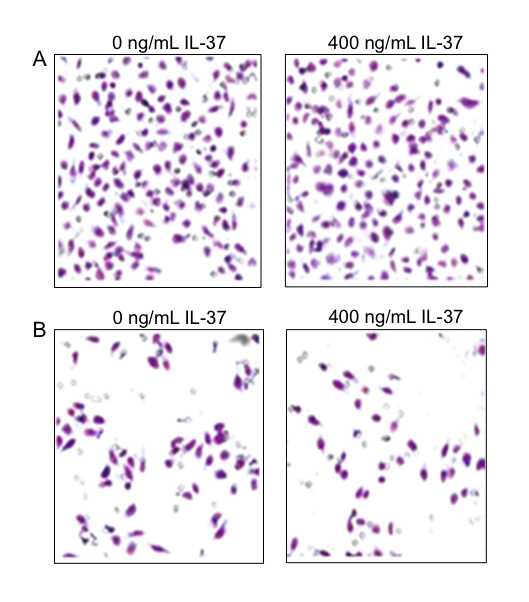 Fig. 1 IL-37 inhibited invasion and migration in HeLa cells.
A) Cell invasion assay. B) Cell migration assay. HeLa cells were treated with 400ng/mL or 0 ng/mL of IL-37 for 72 hrs.
The phosphorylation of STAT3 proteins and the expression of MMP-2, and MMP-9, proteins in IL-37-treated HeLa cells was measured using Western blot. Results showed that phospho-STAT3 and total MMP-2 and MMP-9 protein levels significantly decreased in HeLa cells after treatment with 400ng/mL of IL-37 for 72 hrs compared to cells treated with 0 ng/mL of IL-37 (p < 0.05, Table 3, Fig. 2).
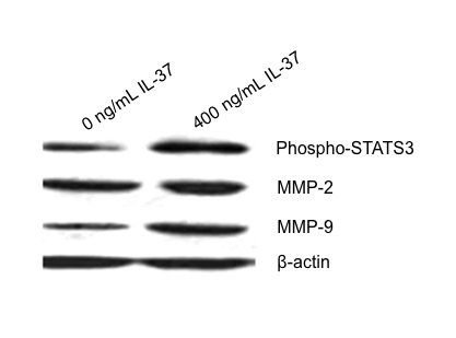 Fig. 2 Western blot of phospho-STAT3, total MMP-2 and MMP-9 expression in HeLa cells.
HeLa cells were treated with 400ng/mL of IL-37 for 72 hrs.
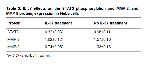 Table 3. IL-37 effects on the STAT3 phosphorylation and MMP-2, and MMP-9 protein, expression in HeLa cells
*p < 0.05 vs. no IL-37 treatment.
The association between cervical cancer, and infection, with human papillomavirus, and other pathogens, has long been known. The inflammatory response resulting from infection is one of primary driving mechanisms of cancer development. The inflammation-induced infiltration of immune cells and their secretory cytokines, chemokines, and growth factors are known contribute to the malignant traits of cervical cancer [12]. This study demonstrated that IL-37, an IL-1 family anti-inflammatory cytokine, significantly inhibited cervical cancer cell proliferation, survival, invasion, and migration. The anti-cancer effect of IL-37 in cervical cancer cells is associated with its ability to decrease STAT3 phosphorylation, and downregulate MMP-2 and MMP-9 protein expression.
Previous studies have shown that IL-37 is expressed in mononuclear and dendritic cells, and weakly expresses in epithelial tissues. IL-37 exerts a strong anti-inflammatory role mainly by suppressing inflammatory cytokine production [13, 14]. IL-18 is a well-known inflammatory cytokine and functions to initiate the inflammatory response and drive Th1, and Th17, inflammatory responses.
Recent studies demonstrated that IL-37 can bind to the IL-18 receptor α chain in a non-competitive fashion and subsequently inhibit IL-18 biological activities. IL-37 also exerts a receptor-independent regulation of inflammation [15]. IL-37’s roles in tumor cells remain relatively unclear. Jiang et al study of cultured renal cell carcinoma cells revealed that IL-37 can serve as a novel tumor suppressor by suppressing IL-6/STAT3 signaling [9]. Zhao et al showed that IL-37 expression decreased in primary hepatocellular carcinoma tissues, which negatively correlated with tumor size and hepatocellular carcinoma patient survival [10]. Wang et al reported that in cultured human cervical cancer cells revealed that IL-37 inhibited STAT3 expression and phosphorylation, which subsequently reduced inflammatory expression of, TNF-α and IL-1β, as well as cell proliferation and invasion [11]. Consistent with previous findings in renal cell carcinoma cells [9] and cervical cancer cells [11], IL-37 significantly inhibited STAT3 phosphorylation in HeLa cells. This study demonstrated, for the first time as far as is known, that IL-37 significantly inhibited MMP-2 and MMP-9 protein expression.
Previous studies have demonstrated that cytoplasmic STAT3 can be activated through phosphorylation at its Ser727 residue. Activated STAT3 can regulate MMP-2 and MMP-9 expression [16, 17]. MMPs could degrade extracellular matrix (ECM), damage basal membrane barrierd, destroy local tissue structures, and facilitate tumor growth, and lead to distal metastasis. MMPs could also induce de novo angiogenesis to provide oxygen and nutrients to tumors for local infiltration and distal metastasis [18, 19]. In this study, Western blot showed that STAT3 phosphorylation and that MMP-2 and MMP-9 protein expression in IL-37-treated HeLa cells significantly decreased, which correlated with a significant decrease in HeLa invasive and migratory abilities. These findings suggest that IL-37 could inhibit cervical cancer cells metastasis by suppressing STAT3- MMP-2 / MMP-9 signal pathways.
Previous studies have demonstrated that IL-37 may decrease the activities of inflammation-related kinases, such as FAK, STAT1 and MAPK during tumor occurrence [20]. STAT3 was frequently found to be overexpressed in tumor cells, and tissues, and to control tumor cell growth through regulating the expression of a number of oncogenic genes [21].
This study did not investigate the changes in the expression of the STAT3-regulated oncogenes, the STAT3 phosphorylation in HeLa cells was down-regulated by IL-37 in a time and concentration dependent manner, which correlated with a reduction in cell viability, arrest of in cell cycle at the G0/G1 phase, as well as an increase in cell apoptosis. STAT3 phosphorylation in HeLa cells was down-regulated by IL-37 in a time and concentration dependent manner. This correlates with reduced cell viability, arrest of SOMETHING in THE cell cycle at the G0/G1 phase, It also correlates with cell apoptosis increase. These findings suggest that IL-37 can inhibit the proliferation and survival of cervical cancer cells possibly by downregulating STAT3 activities.
In conclusion, IL-37 is a tumor suppressor in cervical cancer cells and can inhibit the cervical cancer cell: proliferation, survival, invasion, and migration. This is accomplished possibly by inhibiting STAT3 phosphorylation and reducing subsequent MMP-2 and MMP-9 protein expression. Further studies should be conducted to address whether IL-37 expression is lost in cervical cancer tumor tissues and whether IL-37 expression correlates with the malignancy of cervical cancer and the prognosis of cervical cancer patients.
1.
2.
3.
4.
5.
6.
7.
8.
9.
10.
11.
12.
13.
14.
15.
16.
17.
18.
19.
20.
21.
Yang X, Wang F, Wang L, LChen L, Shi Y. IL-37 Inhibition of Cervical Carcinoma Cell Proliferation and Infiltration. Med One. 2016; 1(1): 1; https://doi.org/10.20900/mo.20160001

Copyright © 2020 Hapres Co., Ltd. Privacy Policy | Terms and Conditions


