Location: Home >> Detail
Regen Med Front. 2020;2(1):e200001. https://doi.org/10.20900/rmf20200001

1 Department of Materials Science and Technology, Nagaoka University of Technology, Kamitomioka 1603-1, Nagaoka, Niigata 940-2188, Japan
2 Center for Functional Sensor & Actuator (CFSN), National Institute for Materials Science (NIMS), 1-1 Namiki, Tsukuba, Ibaraki 305-0044, Japan
3 World Premier International Research Center Initiative (WPI), International Center for Materials Nanoarchitectonics (MANA), National Institute for Materials Science (NIMS), 1-1 Namiki, Tsukuba, Ibaraki 305-0044, Japan
4 John A. Paulson School of Engineering and Applied Sciences (SEAS), Harvard University, 9 Oxford Street, Cambridge, Massachusetts 02138, USA
* Correspondence: Motohiro Tagaya, Tel.: +81-258-47-9345.
This article belongs to the Virtual Special Issue "Multifunctional Biomaterials for Regenerative Medicine"
There has been an increasing demand for the development of cell-labeling nanomaterials that safely label and visualize a specific type of cells for diagnosis and inspection in vivo and in vitro. In order to design such cell-labeling nanomaterials, the properties of efficient visible light luminescence and effective interactions with cells have to be realized using a biocompatible nanomaterial. From this viewpoint, we summarize and overview the current situation on cell-labeling technologies. Among various functional nanomaterials, we focus on hydroxyapatite nanoparticles and their photofunctionalization based on the properly designed inorganic-organic hybrid structure such as hydroxyapatite/organic europium (III) complex. Also, the immobilization technique of a specific binding molecule to the solid surface is introduced to demonstrate the selective uptake into cancer cells. Moreover, an example of the growth inhibitory drug molecules for cancer cells are described, focusing on the cytostatic inhibition of citric acid and the potential use of hydroxyapatite/citric acid hybrids. Finally, we mention our future perspectives on the theranostic nanoparticles with fluorescence and therapeutic properties that are achieved through the hydroxyapatite-organic hybrid interfacial interactions.
Pathological diagnosis and noninvasive therapy have been extensively investigated so far by means of a variety of techniques. Among them, nanomaterials that can bind to the surface of cells and fluoresce by light irradiation with a specific wavelength are well-known as cell-labeling nanomaterials and have been widely utilized for various purposes [1–4]. Many different types of cell-labeling technologies have been developed for the diagnosis of cancer cells including radioisotopes for single photon emission computed tomography (SPECT), positron emission tomography (PET) [5], iron-nanoparticles for magnetic resonance imaging (MRI) [6], and antibody-mediated labeling with optical probes [7]. However, the application of these techniques to early-stage cancer cells is still a grand challenge in this field. In order to overcome various difficulties, one of the keys would be to develop diagnostic techniques using a fluorescence endoscope to detect the epithelial cancer cells.
Other important features that are demanded for the cell-labeling nanomaterials are that they can safely label and visualize the cancer cells for diagnosis and inspection in vivo and in vitro. To realize such cell-labeling nanomaterials, nanomaterial itself has to be biocompatible, efficiently fluoresce in the visible light range (400–800 nm) as well as effectively interact with the cells. For the early stage detection of the cancer cells in vivo, the surface layer of the cells is stained to be observed with a fluorescence endoscope. Then, the cells are diagnosed on the basis of autofluorescence which is known as a fluorescence emitted by cell constituents. A previous study reported that clear luminescence peaks were observed at approximately 500 nm and 630 nm when excited at a wavelength of 437 nm [8]. However, the luminescence intensity of autofluorescence is too weak to confirm with our naked eyes, limiting practical use of this phenomenon to various applications. For this reason, many researchers have started from in vitro studies to achieve luminescence that can sufficiently be observed with a fluorescent endoscope, probably leading to the cell diagnosis in vivo afterwards. Although the detection limit of a confocal laser scanning microscope in z direction is known to be approx. 500 nm [9], some holographic microscopes can observe almost 30 m in depth from the cellular top surface. Taking advantage of these techniques, the cell-labeling nanomaterials can be utilized for immunoassay to diagnose early stage cancer in the visible light region.
To further develop and optimize nanomaterials with desired cell labeling properties, a possible mechanism on how the nanomaterials are captured by cells is considered in relation to the basic structure of cells. Endocytosis is known as one of the representative processes in which the nanomaterial is taken into the cell from the outside via the cell membrane [10]. There are two types of endocytosis: phagocytosis and pinocytosis Phagocytosis is a mechanism that cells take up foreign substances (bacteria, viruses, parasites) and abnormal metabolites into the cell (tissue, blood, etc.) and decompose them. Phagocytosis occurs only in a specific type of cells such as phagocytic cells, monocytes, dendritic cells, NK cells and neutrophils. Pathogens, dead cells and cell debris (1 µm or more) are also included. On the other hand, pinocytosis refers to the phenomenon that cells take up nanomaterials as vesicles regardless of their permeability. Internalization of substances with the size less than 300 nm [11] and 50 to 80 nm [12] into cells have been reported. No clear consensus has been obtained so far with respect to the mechanism of nanomaterial uptake into cells. The surface physical properties are regarded as one of the important factors, while the uptake behavior also varies depending on the types of cells. Since proteins and nanomaterials in vivo bind to each other and agglutinate, the cellular uptake mechanism does not depend only on the size of the nanomaterials. Pinocytosis depends on the size, composition and structure of the nanomaterial surface.
In this review, we summarize and overview some aspects and future challenges of the cell-labeling techniques. The cell-labeling techniques have the potential to be improved by the development of cytocompatible photoluminescence probes [13,14]. For example, lanthanide (Ln) ion-doped nanomaterials provide several advantages such as lower cytotoxicity, photostability and sharp luminescence bands over the conventional cell-labeling nanomaterials including organic molecules and semiconductor quantum dots [15,16]. From this viewpoint, we describe the importance of photofunctionalization of hydroxyapatite, which is realized by designing the inorganic-organic nanohybrid structures. We also highlight the surface immobilization technique of nanomaterials, making it possible to control uptake into cancer cells.
Figure 1 shows an outline of the labeling mechanism (endocytosis). On the basis of the interaction between a nanomaterial surface and cell membrane (receptor), endocytosis is classified into two behaviors: passive and active. The former is a weak liquid phase interaction such as hydrogen bonding, a hydrophobic effect and van der Waals force. In such cases, the nanomaterials are easily taken into the cells since surface energy is same as that of cell surfaces. The latter is a strong liquid phase/solid phase interaction such as ionic bonding, metal bonding and covalent bonding, etc. In the case of the strong interaction, the catalytic effect at the interaction point inhibits cellular uptake because of the high surface tension. The nanomaterial taken up by the cell is carried to the lysosome, then hydrolyzed and digested by enzymes. Likewise, the cell-labeling nanomaterial is taken into the cell through the interaction with the receptor present on the surface of the cell membrane, and fluorescence is emitted by the excitation of visible light. The luminescence emitted from cells in vitro can be detected with a fluorescence microscope and used for diagnosis. Similarly, in vivo, it is detected with a fluorescence endoscope and used for diagnosis under the visible light excitation.
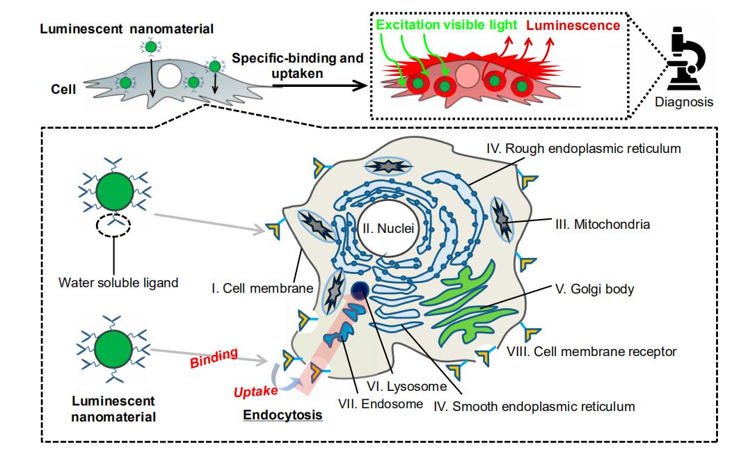 Figure 1. Illustration of the possible cell-labeling mechanism for diagnosis. The site to be taken into the cell is different depending on the particle size as described in the text.
Organic molecules
Organic molecules and inorganic nanoparticles have been used as cell-labeling nanomaterials. Depending on the nanomaterial, there are some problems such as fast color degradation which is due to ultraviolet light excitation, damaging the living tissue (influence of active oxygen generated by photochemical reaction, damage of biomolecules due to electron transfer reaction, etc.). A variety of nanomaterials have been synthesized so far; however, there are still no suitable nanomaterials to solve such problems. Some examples of cell-labeling nanomaterials already reported and known problems are shown in Table 1. For the organic molecules, a fluorescent protein (excitation wavelength: 400 nm) [17], fluorescent organic small molecule (excitation wavelength: 370 nm) [18], temperature responsive polymers (excitation wavelength: 456 nm) [19] and pH responsive dye (excitation wavelength: 532 nm) [20] have been reported. Such cell-labeling nanomaterials are not cytotoxic and do not affect cell function and cell growth behavior. Fluorescent proteins are less toxic to living bodies and emit luminescence through the visible light excitation. However, the protein can be decomposed by the light irradiation, affecting the function of cell-labeling. In the case of fluorescent organic low molecules, their fluorescence properties are affected by the molecular size which frequently causes steric hindrance. Although no such drawbacks are seen with the temperature responsive and pH responsive dyes, some other difficulties including water insolubility and aggregate formation limit their usage in cell-labeling applications. For this kind of applications, it is necessary to synthesize nanomaterials which have sufficient stability in their color and luminescence properties even under continuous/strong light excitation.
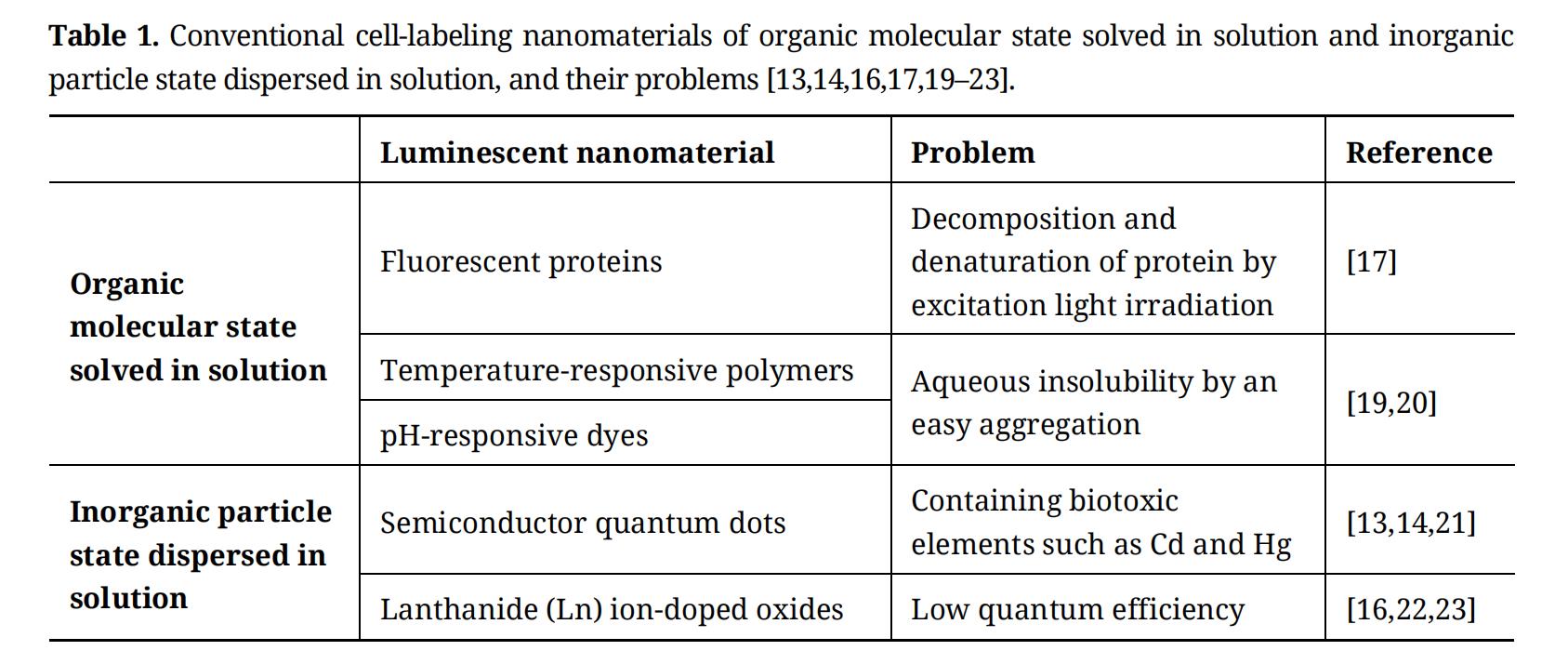 Table 1. Conventional cell-labeling nanomaterials of organic molecular state solved in solution and inorganic particle state dispersed in solution, and their problems [13,14,16,17,19–23].
Inorganic nanoparticles
Among various inorganic nanoparticles, semiconductor quantum dots (excitation wavelength: 260 nm) have been mainly utilized for cell-labeling applications [21] because of their strong luminescence and stable color. However, a problem has been also reported in terms of biotoxicity which is induced by compositional Cd, Hg, etc. (biotoxic element) [13,14,21]. These heavy metals are known to affect the cell function and reduce cell viability, possibly leading to undesirable and/or unexpected results in in vivo experiments. In addition, since the quantum dots are usually excited by UV light irradiation, potential damage to the cells is also concerned. To solve this problem, a Ln ion-doped oxide which is excited by the light with longer wavelength has been reported as the cell-labeling nanomaterial [16,22,23], while its luminescent properties and safety to cells are unknown yet (Table 1).
In order to overcome these barriers, a Ln ion-doped hydroxyapatite (HA) would be one of the model nanomaterials. HA has an excellent affinity with cells so that it can be used as a cell label. It is possible to synthesize cell-labeling HA-based nanomaterials with an excellent cell affinity by doping Ln ions that have low toxicity to living bodies and are capable of being excited by a low energy light in the visible range. Needless to say, the luminescent species (Ln ions) have to be incorporated into HA structure in such a way that their amount, spatial distribution and so on are controlled.
Features
The HA (Ca10(PO4)6(OH)2) with a Ca/P ratio of 1.67 is an inorganic component contained in living tissues (bones and teeth), the crystal system is hexagonal, the space group is P63/m, and the unit cell is 0.94 nm × 0.94 nm × 0.68 nm [24]. As a crystal structure, the four columnar Ca (i.e., Ca (I) site) is aligned parallel to the c axis, and the six screw axis Ca (i.e., Ca (II) site) surround the c-axis. Also, hydroxyl groups are present in the part surrounded by the screw Ca. The crystal structure of HA is shown in Figure 2. The high biocompatibility due to having a composition close to the hard tissues in vivo is an advantage, and applied study as a biomaterial is proceeding. The reported particle size of HA is around 20–300 nm [25,26], and it has excellent light resistance. The uptake of the HA nanoparticles with a size of 50–200 nm by the osteoblasts has been investigated, exhibiting the excellent uptake efficiency without cytotoxicity [27]. The Ca ions in the HA can be easily substituted with the other metal ions, and by substituting Ln ions, a luminescence property can be imparted. By using the HA matrix, it is possible to achieve the cell-labeling nanomaterials that are safe for the living body and do not affect the color degradation and luminescence properties due to the light excitation. Furthermore, since it is possible to impart the other functions (e.g., drug delivery carrier) for achieving the multi-functional particles, it is important to design the cell-labeling nanomaterials as nanoparticle shapes.
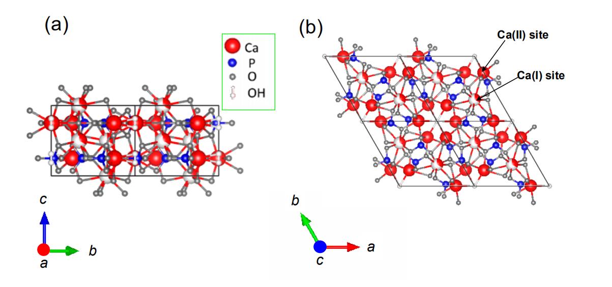 Figure 2. Crystalline structures of HA viewed from the (a) a- and (b) c-axis directions.
Hybrid systems with organic molecules in vivo
Figure 3 shows examples of inorganic-organic hybrids. Figure 3a,b shows the hierarchical structure of the chiton. The chiton is known to concentrate as much as 100,000 ppm of iron in teeth. In general, although organisms often use calcium as the main component when forming hard tissues such as teeth, there are few organisms that form teeth with magnetite (Fe2O3) as the main component. The hydroxyl group of lepidochrocite (α-, γ-FeOOH) is exposed on the surface, and exists as an inorganic-organic hybrid by interacting with α-chitin and proteins [28–30]. This inorganic-organic hybrid is achieved by the chemical bond of the Fe2+ ion of α- and γ-FeOOH with the carboxyl group and hydroxyl group in the α-chitin and proteins.
The bone tissue in vivo is also the same. Figure 3c,d is a schematic diagram of bone tissue in vivo. HA is the main component of bone in vivo, and it forms a chemical bond at the interface with collagen and exists as the inorganic-organic hybrid. This hybrid was achieved by the chemical bonding of Ca ions in HA and carboxyl groups in collagen fibril on the interface. The action of organisms to form minerals is called biomineralization and it is known to also occur in vivo.
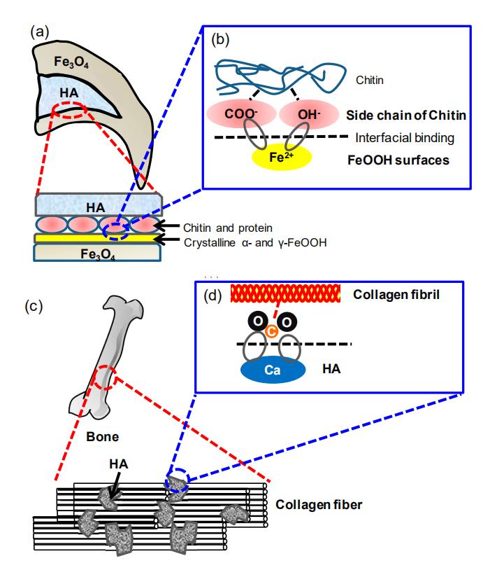 Figure 3. (a, c) Hieratical structures of inorganic-organic hybrids at (a, b) teeth of chiton and (c, d) cancellous bone tissue, and their (b, d) interfacial interactions in the binding state at the molecular level.
The study on the synthesis of functional nanoparticles has been conducted using biomineralization [31,32]. The possibility of the development of the novel cell-labeling nanoparticles by mimicking biomineralization is expected.
Incorporation of Hydroxyapatite Nanoparticles into CellsThe interaction between cytophilic nanoparticles and cells depends on the particle size, surface structure, chemical composition, solubility and aggregation. These are important for circulation in the body, invasiveness to lesions and accumulation, and can be expected to improve the effects of diagnosis and therapy.
The substances diffuse into the cells due to the concentration gradient. The transport of the substances inside and outside the cell membranes by the concentration gradient without chemical change is called as passive transport [33]. In the passive transport, the substance is transported by the concentration gradient. On the other hand, transporting the substances against the concentration gradient is called as active transport [34]. The active transport is performed by a pump in the cell using the hydrolysis energy of adenosine triphosphate. However, the selective intracellular targeting is difficult to be done using these intracellular transports. Therefore, we have been focusing on the transport through endocytosis, and the selective cellular uptake can be achieved by designing the surface immobilization states of the nanoparticles. Since several endocytosis-mediated cellular uptake mechanisms have been proposed, the size of the nanoparticle to be incorporated is also an important factor in cellular uptake. As previously mentioned, the uptake of HA nanoparticles with a size of 50–200 nm (100 μg/mL) has been reported in osteoblasts [35]. In particular, the particle size of about 80 nm for HA particles has been reported to be good for cellular uptake. Specifically, the cells taking up the HA nanoparticles showed the same growth behavior as non-incorporated cells, and the cell affinity with the particles alone was confirmed. It has already been defined that there is toxicity to cells when the cell growth rate is less than 130% in 6 h of culture [36]. As an international standards, the nanoparticle is defined as non-cytotoxicity, if the decrease rate in the cell viability during the cell culture is 30% or less as compared with the case in the cells without the nanoparticle [37]. The HA particles can be judged to be non-cytotoxic in the size range of 50–200 nm [35] according to previous reports [36,37]. For the cell-labeling, the particle sizes of HA in a media bound to the cells via endocytosis was approximately 120 nm [38]. Thus, HA is suitable as the matrix for the cell-labeling nanoparticles.
Possibility of Imaging by PhotofunctionalizationHybrids with functional low molecular compounds such as 8-hydroxyquinoline (8Hq) [39,40] and glucosamine [41] on a nanoscale have already been reported. For example, in the HA/8Hq system, the 8Hq molecules were chemically bonded to the Ca2+ ions on the HA by applying mechanical forces. The photofunction of the hybrids has been studied by evaluating the light absorption and luminescence through the metal-ligand charge transfer complex formation. In the HA/glucosamine system, glucosamine molecules were hybridized with HA due to the hydrogen-bonding interactions between the amino and hydroxyl groups of glucosamine and the hydroxyl groups on the surface of HA. No study has been done yet to hybridize a photofunctional dye (visible light responsive) and HA and use them as cell labels. Several studies of the europium (III) ion (Eu3+) doped HA have been reported [42,43]. For example, when the Eu3+ doped HA was synthesized in the presence of cetyltrimethylammonium bromide as a cationic surfactant, the substitution efficiency of the Eu3+ ion into the Ca (II) site of HA was enhanced [44]. However, the low quantum yield is a problem when compared to existing the cell-labeling nanoparticles.
Thus, detailed understanding of the photophysicochemical driving force at the interface with organic molecules on the HA surface is considered to be important for the design of novel cell-labeling nanoparticles.
For the Ln ions, most of the absorption and luminescence transitions in the infrared to near-ultraviolet region are due to transitions between the split 4f orbital levels [45,46]. The electric dipole transition between energy levels belonging to the same electron configuration becomes a forbidden transition by the selection rule of evenness and oddity. However, when atoms or ions are in a solid or in a liquid, the 5d level on the 4f level mixes with the 4f level due to the effect of the crystal field, resulting in an allowable transition. Even in the case of the crystal field having an inversion symmetry, it is permitted to some extent by coupling with the lattice vibration having odd properties. The transition probability changes depending on the symmetry of the ligand field and the electronic state of the Ln ion. The 4f orbitals of the Ln ions are shielded by the 5s and 5p orbitals [47], which increases the fluorescence lifetime. The six electrons in the 4f shell can be arranged into seven 4f orbitals. The degeneracy of a 4fn electronic configuration is given by the binomial coefficient using the following Equation (1).

where n is the number of 4f electrons. The n of Eu3+ is 6. The energy diagram of the Eu3+ ion is shown in Figure 4. All the peaks corresponded to the transitions from the metastable orbital singlet state of 5D0 to the spin–orbital states of 7FJ (J = 0, 1, 2, 3, 4) of the Eu3+ ion. J represents the total angular momentum. These characteristic transitions were assigned to the 5D0 to 7F0 at about 575 nm, 7F1 at about 590 nm, 7F2 at about 616 nm, 7F3 at about 653 nm and 7F4 at about 698 nm, respectively. In particular, the transitions of 5D0 to 7F1 are called magnetic dipole transitions, and the transitions of 5D0 to 7F0, 2, 3, 4 are called electric dipole transitions. In the case of Eu3+, the electric dipole transition from the excited state 5D0 to the 7F0, 2, 3, 4 level is forbidden if the substitution site has an inversion symmetry. On the other hand, it is known that the occurrence probability of a magnetic dipole transition is irrelevant to symmetry. When an atom or crystal is in the molecule, electrons belonging to the atom of interest undergo a Coulomb interaction from the charge in the surrounding ions. The Coulomb potential due to such a surrounding charge is called the crystal field potential. Due to this crystal field potential, the energy levels of atoms that degenerate in the free atom state split. The degeneracy of the 4f6 configuration is partly or totally lifted by several perturbations acting on the Eu3+ ion; i.e., electron repulsion, spin–orbit coupling, the crystal-field perturbation and eventually the Zeeman effect. The electron repulsion is the electrostatic interaction between the different electrons in the 4f shell. The spin–orbit coupling results from the interaction between the spin magnetic moment of the electron and the magnetic field created by the movement of the electron around the nucleus. The crystal-field effect is caused by the interactions between the 4f electrons and the electrons of the ligands. The Zeeman effect is the splitting of the energy levels by an external magnetic field.
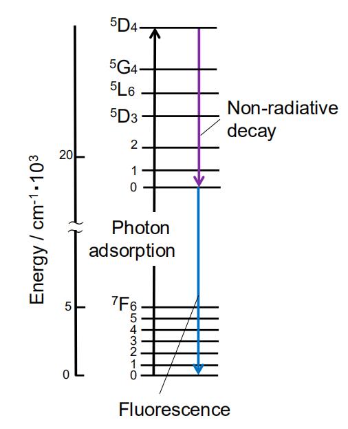 Figure 4. Energy diagram of Eu3+ and the possible excitation and decay processes. The excitation processes consist of 7F0→5D4, 7F0→5G4, 7F0→5L6, 7F0→5D2 and 7F0→5D1, and the decay processes corresponded to the transitions from the metastable orbital singlet state of 5D0 to the spin–orbital states of 7FJ (J = 0, 1, 2, 3 and 4) of Eu3+.
The splitting of the terms into the J states by the spin–orbit coupling interaction is on the order of 1000 cm−1. The 2J + 1 degeneracy of the energy levels in the free ion is further lifted by the crystal-field effect, after which the energy levels are characterized by the irreducible representation of the point group of the Eu3+ site [48]. These levels are called crystal-field levels (or Stark levels). The splitting of the energy levels by the crystal-field effect is on the order of a few hundred cm−1 or less. In systems with an orthorhombic or lower symmetry, all the degeneracy are lifted by the crystal field. In systems with a higher symmetry, all of the degeneracy can be lifted by an external magnetic field, via the so-called Zeeman effect. Even in strong magnetic fields, the splitting of the energy levels by the Zeeman effect is only a few cm−1. The J quantum numbers are well defined in the free Eu3+ ion, but J-mixing occurs when the Eu3+ is located in a non-spherically symmetric ligand environment [49]. J-mixing is induced by the even-parity components of the crystal-field potential.
The energy levels and wave functions of the Eu3+ ion can be obtained by diagonalization of the energy matrix [48]. The matrix elements are of the type <Ψ|H|Ψ′>, where H is the effective-operator Hamiltonian, and Ψ and Ψ′ are basis functions of the 4fn configuration (n = 6 for Eu3+). The angular parts of the matrix elements can be exactly calculated, whereas the radial parts are treated as adjustable parameters. A parameter set is obtained by optimizing a start set of parameters by a general least-squares fitting process in which the energy differences between the calculated and experimental energy levels are minimized. The total Hamiltonian can be written as the sum of a free-ion and a crystal-field part by the following Equation (2).

The free-ion Hamiltonian is characterized by a set of three electron repulsion parameters (F2, F4, F6), by the spin–orbit coupling constant ζ4f, the Trees configuration interaction parameters (α, β, γ), the three-body configuration interaction parameters (T2, T3, T4, T6, T7, T8) and parameters which describe magnetic interactions (M0, M2, M4, P2, P4, P6). An additional parameter Eave (ave stands for “average”) takes into account the kinetic energy of the electrons and their interaction with the nucleus. The free-ion Hamiltonian can be written by the following Equation (3) [50,51]:
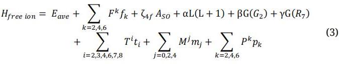
where fk and ASO represent the angular part of the electrostatic and spin–orbit interaction, respectively. L is the total orbital angular momentum. G(G2) and G(R7) are the so-called Casimir operators for the groups G(G2 and R7, respectively. The ti are the three-particle operators. The mj and pk represent the operators for the magnetic corrections. The Fk parameters decrease if k increases. The terms in the Hamiltonian that represent the non-spherical part of the interactions with the host matrix are described by using the crystal-field Hamiltonian. The crystal-field Hamiltonian can be written by the following Equation (4) [48]:

where Cq(i) are tensor operators of rank k with components q. These tensor operators transform the spherical harmonics. The Bq is the crystal-field parameter, n is the number of electrons (6 in the case of Eu3+) and i represents the i-th electron. The number of non-zero parameters are determined by the point-group site symmetry of the Ln ion. For cell-labeling applications of the Eu3+ ions, it is important to change the symmetry and enhance the luminescence.
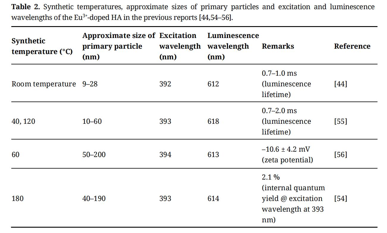
There are many ions in vivo that play important roles in the expression of various biological functions. The HA can be given various functions by the substitution of ions in the crystal structure. The studies of ion substitution in the HA crystal structure have been conducted [52–54]. In particular, it is known that the Ca2+ can be substituted by various metal ions. Table 2 shows some reports on the doping of Eu3+ ions into the HA structure which is considered to be useful as a cell-label. Among the Ln ions, the Eu3+ ion is known to have a low cytotoxicity. The synthesis temperature has been controlled to synthesize the Eu3+ ion-doped HA. Excitation and luminescence spectra measurements were performed in the range of the excitation wavelength of 392–394 nm and luminescence wavelength of 612–618 nm [44,54–56]. The internal quantum yield, fluorescence lifetime and zeta potential were reported as other evaluation remarks in the references. The fluorescence lifetime of about 0.7–2.0 ms was achieved [44,55], the zeta potential of about –10.6 mV was reported [56], and the internal quantum yield of 2.1% was achieved by the excitation light irradiation of 394 nm [54]. Figure 5 shows a diagram of the coordination structure of oxygen to the Ca site in HA. The Ca (I) site has a coordination number of 9, and C3 symmetry, and the Ca (II) site has a coordination number of 7, and Cs symmetry. The transition from 5D0 to 7F0 is known to indicate the presence of Eu3+ ions at the Ca (I) and Ca (II) sites in HA. The 5D0 to 7F0 transition peak appears at a maximum fluorescence wavelength of 572 nm at the Ca (I) site and 577 nm at the Ca (II) site [44]. By separating the peak of these two wavelengths, it is possible to evaluate the ratio of Eu3+ ions present at the Ca site. The photofunctionalization of HA using the Eu3+ ion is the subject of study. However, due to the low internal quantum efficiency, almost no study has been reported on its application in the cell labeling. Thus, the state of Eu3+ ions in the HA is important regarding the luminescence behavior.
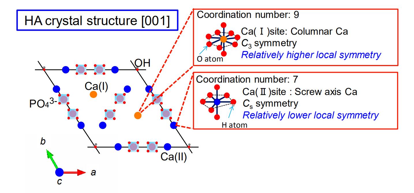 Figure 5. Coordination states of Ca (I) and Ca (II) sites in the HA structure.
Types and photochemical properties
As functional low molecular weight compounds, the Eu (III) complexes having a ligand are interesting because of their luminescent properties and have been studied in the biofield and the electronics field. The Eu (III) complexes have characteristic narrow luminescence spectrum lines and long-lived excited states. The ligands effectively increase the light absorption. Although the spectral shape depends on the electronic environment, due to the spatially shielded f-f transition, the Eu3+ ion alone has a low light absorption ability. It is known that a strong luminescence is observed in the Eu (III) complex in which various ligands are bound to the Eu3+ ion. The ligand absorbs the ultraviolet light, and the energy is transferred to the Eu3+ ion and finally observed as the luminescence. The effect of ligand is called the antenna effect. When electrons in a singlet ground state molecule are excited to a high energy state by light absorption, they become an excited singlet state (S1) or an excited triplet state (T1). The S1 is a molecular state in which all the electron spins are paired, and the spins of excited electrons are become the opposite direction to the ground state electrons. In the T1, the spins of the excited electrons become the same direction as that of the ground state electrons. Since the excitation to the T1 includes the inversion of spin, which is a forbidden transition, the probability of the molecule forming the T1 by light absorption is low. The processes of non-radiatively changing from the S1 to T1 or T1 to S1 are called intersystem crossing. When the vibrational levels of the two excited states overlap, the probability of intersystem crossing increases because the change in energy due to the transition is low. Figure 6 shows examples of Eu (III) complexes that have been reported for cell-labeling. Some β-diketonato Eu (III) complexes have been used for the cell-labeling [57,58]. These reports describe that the Eu (III) complex was dissolved in phosphate buffered saline, then cell-labeling was performed. In the case of the β-diketonato Eu (III) complex of Figure 6a, the excitation/luminescence spectrum was measured at the excitation wavelength of 402 nm and the luminescence wavelength of 614 nm, and the internal quantum yield was 41% [57]. In the case of the β-diketonato Eu (III) complex of Figure 6b, the excitation/luminescence spectrum was measured at the excitation wavelength of 405 nm and the luminescence wavelength of 612 nm, and the internal quantum yield was 23% [58]. However, these available Eu (III) complex luminescent probes have the problem of color degradation, when the probes are exposed to a continuous and intense excitation light for monitoring the biological processes. By introducing an asymmetric coordination field into the Eu (III) complex, the forbidden transition is broken to allow the f-f transition, and the light absorption coefficient can be increased to improve the luminescence intensity. If the symmetry of the ligand can be lowered, application as a cell-labeling probe can be expected.
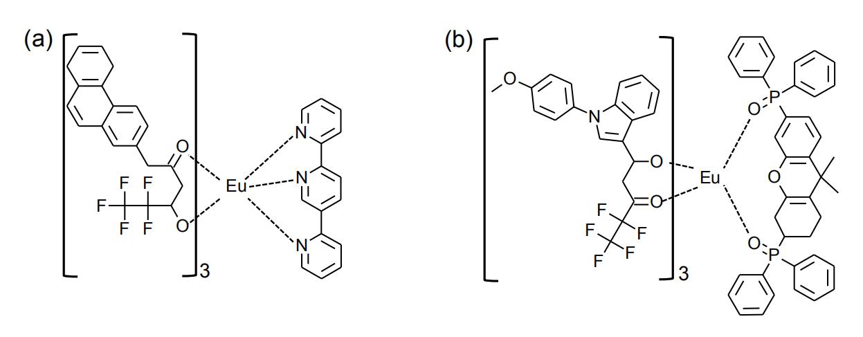 Figure 6. Eu3+ complexes of (a, b) β-diketonate ligand [57,58]. Internal quantum yield of (a) is 41% (λex = 402 nm, λem = 614 nm), and (b) is 23% (λex = 405 nm, λem = 612 nm). The Eu3+ complexes have been studied for biological labelling applications.
Tris(2,2,6,6-tetramethyl-3,5-heptanedionato) europium (III)
The molecular model of tris(2,2,6,6-tetramethyl-3,5-heptanedionato) europium (III) (EuTH) as a complex having the diketonato ligand is shown in Figure 7. This complex has a highly-symmetrical molecular structure, and is insoluble in water but soluble in ethanol. Due to the high symmetry of the ligand, the luminescence intensity is low.
In the EuTH, it is known that the distance between the Eu–O and the ligand and the coordination structure affect the electron transition [59]. Different electronic transition states are observed by the coordination symmetry structural change, and the electric dipole transition is strongly observed in the luminescence spectra. Since it can be dissolved in ethanol, the synthesis of a homogeneous hybrid with HA can be expected.
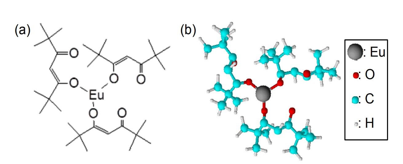 Figure 7. Chemical structures of EuTH molecule at the (a) 2- and (b) 3-dimensional views.
Possibility of hybrid synthesis of hydroxyapatite with EuTH
Ln complexes incorporated into an inorganic porous matrix, such as zeolite or mesoporous silica, that forms an inorganic-organic hybrid have been reported [60,61]. The confinement of the Ln complex within the inorganic porous structure not only improves its stability but also reduces the quenching due to aggregation between the Ln complexes. However, the hybrid with luminescent properties based on the inorganic-organic interface of highly biocompatible HA nanoparticles has not been reported. It is expected that the electron localization between the ligand of the Ln complex and the central metal can be used as a nucleation site of the HA crystals. It was also possible that a high luminescence behavior can be achieved. Specifically, we have been focusing on the electron localization between the central Eu3+ ion of EuTH and the diketonate ligand. The nucleation of HA in the electron localization of EuTH resulted in the synthesis of the HA-EuTH hybrid [62,63]. It is suggested that the luminescence intensity can be improved by lowering the symmetry of the EuTH ligand.
Receptors are present in all cells. The receptor is a substance and is present on the cell membrane, which specifically binds to a substance outside the cell membrane and is usually a protein. Cells can identify specific substances by receptors and receive information from outside the cells. Binding of the receptor to the substance triggers a variety of responses in the cell. Table 3 shows the types of receptors expressed on the surface of epithelial cancer cells that can be treated with an endoscope [64–69].
The types of overexpressed receptors differ depending on the type of cancer. It is known that specific recognition of these receptors enables particle uptake into the cells.
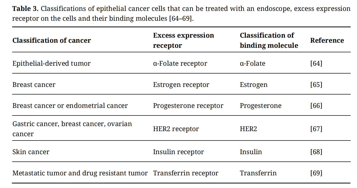 Table 3. Classifications of epithelial cancer cells that can be treated with an endoscope, excess expression receptor on the cells and their binding molecules [64–69].
Table 3 also shows the molecules that recognize and specifically bind to the receptors. The recognition of organic molecules or proteins corresponding to the receptor allows the particle uptake into the cells. In order to be used as the cell-labeling nanoparticles, the molecules should be present in the dispersed state on the particle surfaces. The aggregation of the molecules may make it difficult for the receptor to recognize the molecules. Thus, the efficient uptake of HA nanoparticles into the cells becomes possible by immobilizing the cells binding molecules on the HA surface.
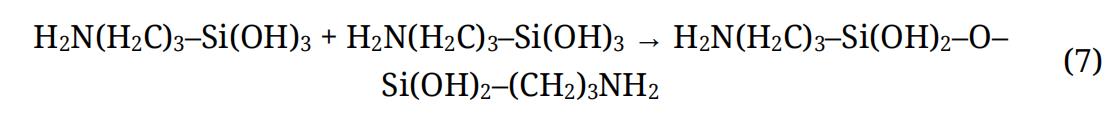
Immobilization Technique of Specific Binding Molecule on Solid SurfaceBy immobilizing a specific binding molecule on a solid surface, it is possible to impart new properties such as in vivo stability, tissue targeting, cell tropism, etc., to the nanoparticle. In order to maximize the properties of the nanoparticles in vivo, it is necessary to achieve surface immobilization techniques that consider the interaction between the cells and particles. The surface immobilization techniques are both chemical (covalent bond) and physical (non-covalent bond). One of the highly-stable chemical methods is reacting a functional group on the solid surface with a specific binding molecule. In this method, since the reactive groups of the molecules immobilized on the surface cause steric hindrance, the polymerization density decreases. Inorganic particles having a hydroxyl group on the surface can be easily made into a functional surface by the method. As an example, hydrophilic amorphous silica particles are negatively charged (≡Si–OH), because the oxygen present in the skeleton of –SiO4– is partially deficient. When the hydroxyl group on the surface of the amorphous silica is reacted with 3-aminopropyltriethoxysilane (APTES, H2N(H2C)3Si(OCH2CH3)3), trimethoxy (2-carboxyethyl) silane (HOOC(CH2)2Si(OCH3)3) and the methylphosphonic acid 3-(trivinylxylsilyl) propyl salt (NaPO3(CH2)3Si(OH)3), etc., it becomes positively or negatively charged by the covalent bond. Specifically, the hydroxyl group of the silicon alkoxide and the hydroxyl group of the solid surface form a hydrogen bond, and the covalent bond is formed by dehydration condensation. Thereby, reactive functional groups, such as amino groups (–NH2) and carboxyl groups (–COOH) can be present on the solid surface. The following is an example of the reaction formula (reaction of APTES).
Formation of silanol group by hydrolysis (5)

Adsorption on the solid surface (6)

Self-assembly reaction of hydrolyzed silanes (7)
where, ● is a solid surface.
Figure 8 shows an example of the immobilization of molecules already reported. Figure 8a shows the immobilization process of APTES on a solid surface. By bonding an amino group-terminated silane coupling agent to the solid surface, the OH group on the solid surface is bonded to Si. Figure 8(b-1,b-2) shows the immobilization processes of molecules on the solid surface. These are examples of the immobilization of a molecule via the APTES [38,70,71]. The following is an example of the reaction formula (8) (reaction of Figure 8(b-1)).

where, H2N–● is an amino group-immobilized solid surface.
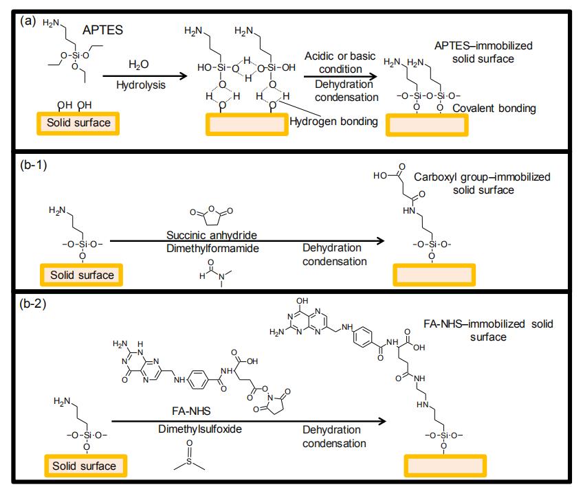 Figure 8. Immobilization processes of (a) APTES, (b-1) carboxyl group and (b-2) FA-NHS on the solid surface. In this case, the representative APTES-immobilized solid surface changed from an amine to (b-1) carboxyl group and (b-2) FA-NHS were shown.
The surface amino group stably immobilizes the molecule through the peptide (–HN–CO–) bond. Specifically, APTES is bonded to the solid surface by a liquid phase reaction, the solid surface is coated with the –Si–O–Si– bond, and the amino group is exposed to the surface. Next, the molecule is formed via a covalent bond to the solid surface by a dehydration condensation reaction (–NH2 + –COOH → –NH–CO–) of an amino group and a carboxylic acid in the molecule. Such a reaction mechanism makes it possible to immobilize folate derivatives (folate N-hydroxysuccinimidyl ester (FA-NHS)), which are known to specifically bind to cancer cells, on the solid surfaces [38]. As the report of the physical surface immobilization technique, the immobilization of ferritin by an electrostatic interaction was achieved on the solid surface [72]. The uptake of the ferritin-immobilized particles into cells was also observed. Thus, for the solid surface, various surface states can be designed.
Table 4 shows examples of cytostatic drug molecules. Vitamin C exerts a strong antioxidant action and generates a large amount of hydrogen peroxide [73]. Although normal cells can neutralize hydrogen peroxide, cancer cells cannot neutralize it, causing cell death. Fucoidan has been shown in basic studies to have the function of transmitting stress signals to the endoplasmic reticulum and inducing cell death [74]. Caffeine has been reported to inhibit cancer cell DNA repair and suppress the growth of cancer cells. It has also been reported that the caspase 9/3 pathway, which is one of the signal transduction pathways causing cell death, functions as a persistent anticancer agent by activation [75]. Citric acid is known to induce cell death by inhibiting the function of proteins involved in cell growth by forming a chelate in the cell with minerals such as iron and calcium [76,77]. It has also been reported that the glycolytic system of the cell is inhibited to suppress proliferation [78]. If these drug molecules can be hybridized with the HA, they may be applied as the nanoparticles for cancer therapy.
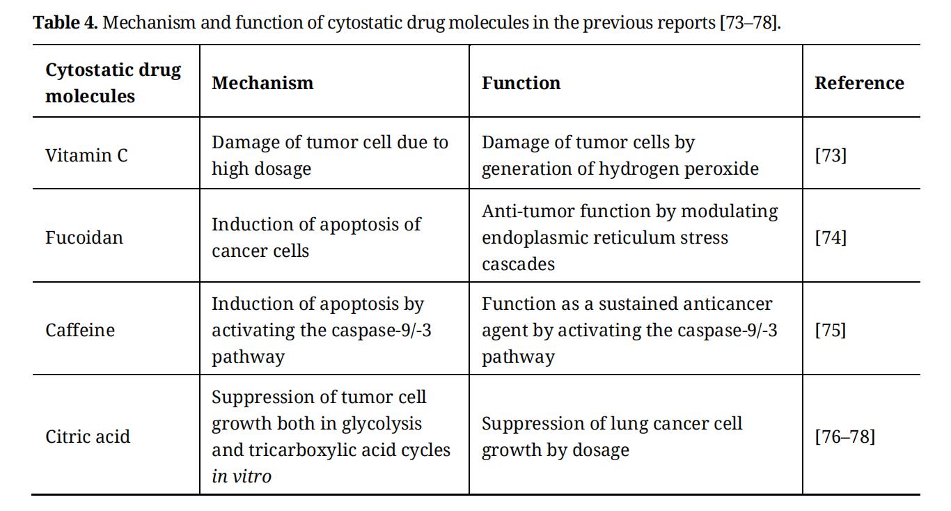 Table 4. Mechanism and function of cytostatic drug molecules in the previous reports [73–78].
Figure 9 shows the molecular structure of citric acid (Figure 9a), dissociation state in solution (Figure 9b) and mechanism of suppression of cancer cell growth by citric acid in the cancer cells (Figure 9c). When the amount of citric acid is increased in the cancer cells, chelates are formed with calcium, iron, copper and zinc, which inhibit the action of proteins and antioxidant enzymes involved in cell growth. As a result, it has been reported to suppress the growth of cancer cells and induce cell death. In the blood, iron (III) ions (Fe3+) bind to transferrin and are transported into the cells. Two Fe3+ ions bind to one transferrin. Transferrin binds to the transferrin receptor located on the cell membrane and is taken up by endocytosis. When it becomes an acidic environment in the lysosome (endosome), Fe3+ ions are then dissociated into iron (II) ions (Fe2+). It then forms the chelate complex with citric acid, depletes Fe2+ ions in the cytoplasm, and inhibits the growth of the cells [79]. The dissociation constant pKFe between citric acid and Fe2+ ion is about 19.7 [80], and the dissociation constant pKCa between citric acid and Ca2+ ion is about 4.3 [81]. In the cells, stable chelates are formed between the Fe2+ ions and citric acid. In normal cells (e.g., prostatic epithelium cells) [82], it is known that the addition of citric acid has no effect on the cellular activity.
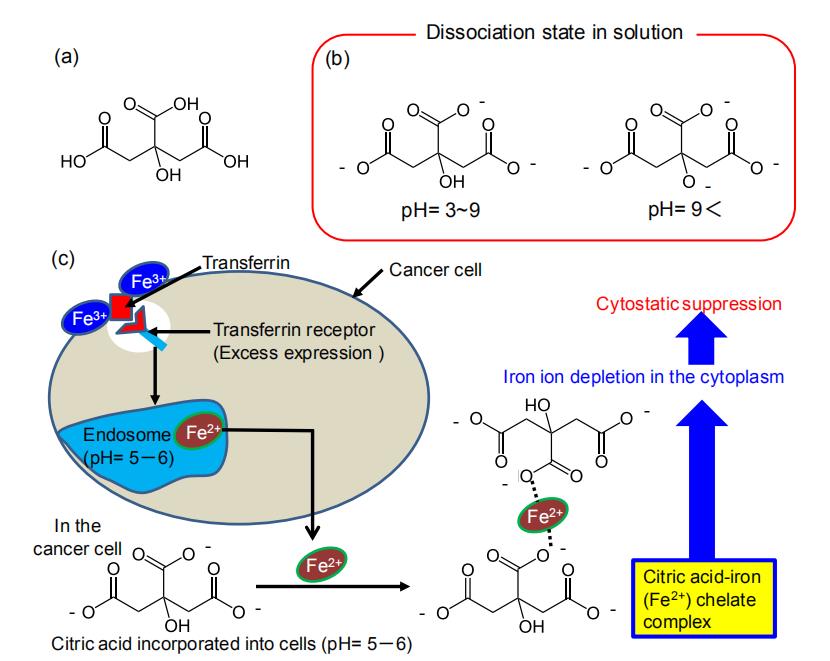 Figure 9. (a) Chemical structure of citric acid and (b) dissociated state in solution at the different pH (pKa1 = 3.12, pKa2 = 4.76, pKa3 = 6.39). (c) Cytostatic mechanism by citric acid. Citric acid and Fe2+ ions can form a chelate complex, and the depletion of Fe2+ ions in the cytoplasm can suppress the cancer cell proliferation. The dissociation constant pKFe between citric acid and iron (II) is about 19.7, and the dissociation constant pKCa between citric acid and Ca2+ is about 4.3. In the cells, stable chelates are formed between Fe2+ and citrate.
Citric acid molecule is used to control the form of HA and is known to exist as the carboxyl ion of –COO- in alkaline solution and as a dimer by hydrogen bonding in the form of (–COOH)2 in an acidic solution [83]. Since HA has Ca sites in the a-axis direction and phosphate sites in the c-axis direction, it has been thought that citric acid molecule adsorbs on the Ca sites and affects the formation of the HA nanoparticles. During HA formation in alkaline solution, it has been suggested that the carboxyl ion of the citric acid molecule and Ca2+ ions of HA chemically bond perpendicular to a-plane (100) on the HA. Citric acid molecule easily forms the chelate with the Ca2+ ions of the HA. On the basis of these results, it has been considered that the competitive reaction between the HA formation and chelate formation enables the synthesis of hybrid nanoparticles. The synthesis of hybrid nanoparticles can be achieved by utilizing the interaction between ions present at the Ca sites in the HA and carboxyl ions of citric acid molecule.
Various types of cell-labeling nanomaterials that stain specific cells have been developed for the non- cytotoxicity visualization. However, two significant problems are known such as color degradation and toxicity in conventional nanomaterials. HA is one of the excellent options to overcome the barriers based on its high biocompatibility; HA is present as a main component of hard tissues in vivo, making it possible to be utilized as a biomaterial. When aiming at the cell-labeling application of HA, it is important to bind a photofunctional molecule with HA in the nucleation process and to synthesize nanoparticles as a form of inorganic-organic hybrid. We have achieved rapid cell-labeling through specific uptake into cancer cells and an easily observable luminescence using the HA-organic complex hybrid. Further studies are now in progress to develop theranostic nanoparticles with fluorescence and therapeutic properties toward future applications in such a field as biomedicine. In order to design novel theranostic nanomaterials, one of the keys would be to control interfacial interactions between HA and organic molecules. To explore the combination of HA and various organic molecules will lead to a large number of multifunctional nanoparticles that advance the frontier technologies of bio-related fields.
The authors declare that they have no conflicts of interest.
This review paper was supported by a grant from the Japan Society for the Promotion of Science (JSPS) KAKENHI (Grant-in-Aid for Young Scientists (A), Grant No. 17H04954, and Grant-in-Aid for JSPS Fellows, Grant No. 18J20271).
1.
2.
3.
4.
5.
6.
7.
8.
9.
10.
11.
12.
13.
14.
15.
16.
17.
18.
19.
20.
21.
22.
23.
24.
25.
26.
27.
28.
29.
30.
31.
33.
33.
34.
35.
36.
37.
38.
39.
40.
41.
42.
43.
44.
45.
46.
47.
48.
49.
50.
51.
52.
53.
54.
55.
56.
57.
58.
59.
60.
61.
62.
63.
64.
65.
66.
67.
68.
69.
70.
71.
72.
73.
74.
75.
76.
77.
78.
79.
80.
81.
82.
83.
Kataoka T, Shiba K, Tagaya M. Design of Hydroxyapatite-Based Multifunctional Nanoparticles for Cell Labelling and Cell Growth Inhibition. Regen Med Front. 2020;2(1):e200001. https://doi.org/10.20900/rmf20200001

Copyright © 2020 Hapres Co., Ltd. Privacy Policy | Terms and Conditions